
Published on the 27th June 2017 by ANSTO Staff
ANSTO and Griffith University environmental researchers and imaging scientists have contributed to a better understanding of how pollutants accumulate and are distributed in amphibians during the larval and metamorphic stages of development.
The investigators, who have published their findings in Environmental Science and Technology, used nuclear techniques to analyse where excess selenium accumulates in tadpoles.
The study showed that tadpoles retained levels of selenium from the larval stage through metamorphosis.
Importantly, the study also demonstrated that accumulated selenium could transfer within the animals as tissues are remodelled or degenerated during amphibian development.
 |
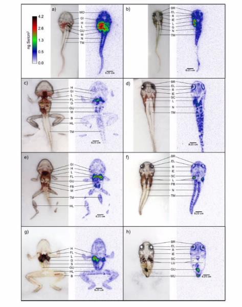
| Autoradiographs reveal biodistribution of selenium throughout metamorphosis |
“The findings may help explain why amphibians in the larval stage have an increased sensitivity to contaminants,” said Dr Tom Creswell, a co-author of the paper with Dr Chantal Lanctôt (below right).
“Chantal has done some outstanding work which has revealed how a clear picture of the biodistribution of a toxic substance is essential to understand all risks associated with exposure.”

Selenium is a macronutrient that is essential for health but excess concentrations bring toxicity.
Anthropogenic activities, such as mining, agriculture and the consumption of fossil fuels have been found to release selenium into surrounding waterways.
Using radioisotopic tracers and autoradiographic imaging at ANSTO, the investigators were able to visually identify the sites in the tadpoles where selenium had accumulated.
Tadpoles are rapidly growing organisms, and they undergo substantial physical and physiological changes as they develop into frogs.
Tadpoles of the native species Limnodynastes peronii were exposed to a low concentration of selenium (in the form of selenite) in water for seven days.
The investigators chose to evaluate a low concentration of selenite, which was within the range found in polluted surface waters, to evaluate bioaccumulation without causing an overt toxicological response from the tadpole.
After seven days, the whole body content of selenium was equivalent to 1.9 μg Se/g dry weight. After transfer to clean water, the tadpoles had eliminated most of the selenium (42% in the first 3 days and a further 41% over the following 10 days). Only 10-14% of the initial selenium remained at the end of the experiment.
The selenium was predominantly found in the liver, kidney, gut and gallbladder. The proportion of selenium in the liver and gallbladder increased 3-4 times during development.
Concentrations were drastically reduced in the excretory organ and gut during metamorphosis.
“The proportion of selenium within the eyes of the tadpole increased during its development and this is an important finding because of the known association of selenium with ocular malformations in other species, including humans,” said Lanctôt.
Audioradiographs revealed that it accumulated predominantly within the ocular lens with lower levels in the retina.
“To our knowledge, this is the first report of selenium accumulation in the eyes of an amphibian, “ said Lanctôt.
The study was an extension of previous research on the same species published in Aquatic Toxicology, which compared accumulation kinetics between two different forms of selenium common to aquatic environments, selenite and selenate, and found that tadpoles accumulated significantly more selenium in the form of selenite.
The research team is now applying Synchrotron-based imaging tools to better understand the accumulation and biodistribution of selenium, and other trace elements, in developing amphibians.
The study was carried out with the assistance of an Australian Institute for Nuclear Science and Engineering (AINSE) Research Award