
Published on the 14th January 2020 by ANSTO Staff
Scientists from the Centre for Crop and Disease Management (CCDM) and Curtin University in Western Australia have used an advanced imaging technique at the Australian Synchrotron for an in-depth look at how a fungus found in wheat crops is damaging its leaves.
Prof Mark Gibberd, director of the Centre, said the investigation was thought to be one of the first that utilised high-resolution X-ray imaging to examine biotic stress related to fungal infection in wheat.

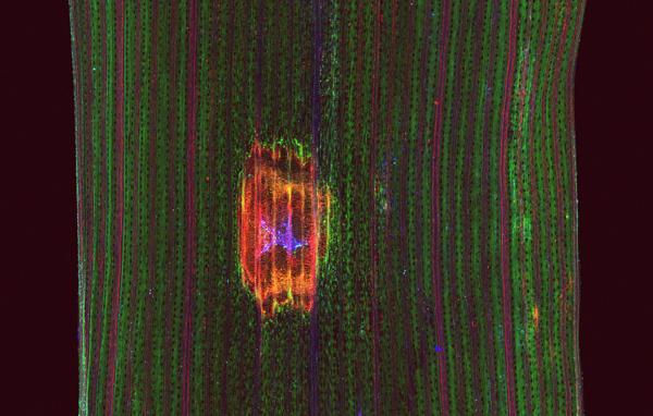
XFM image reveals elements present in yellow spot fungs and the wheat leaves. Image courtesy of Curtin University
Using X-ray fluorescence microscopy (XFM) on leaf samples collected from wheat plants, the team, which included project leader Dr Fatima Naim and ARC Future Fellow Dr Mark Hackett, mapped specific elements in the leaves in and around points of infection.
“Our research project looks at the physiological impact of plant diseases, such as yellow spot, on the function of leaves,” said Gibberd.
Yellow spot is a ubiquitous fungal disease caused by Pyrenophora tritici-repentis (Ptr). It can reduce grain yields by up to 20 per cent – a significant amount which could be the difference between a profitable and non-profitable crop for a farmer.
In Australia, it is one of the most costly diseases to the wheat industry, with wheat yield losses due to yellow spot estimated at over $210 million per year.
The fungus produces effectors, which are toxins that induce necrosis (cell death) in sensitive host varieties.
"While we continue to learn more about the host plant interactions and the role of effectors we also know that the impact of infection on photosynthesis extends well beyond the areas displaying visible symptoms."
“We are confident that the disease is impacting on a much greater leaf area than visible lesions.
The use of X-ray imaging allows us to examine these areas in very high resolution. We plan to compare our findings from the x-ray imaging with information from other techniques including multi-omics approaches and chlorophyll fluorescence imaging to evaluate photosynthetic performance,” said Gibberd.
Breeding methods can produce strains that reduce its impact “We are really excited to be using XFM to help us understand what is happening at the tissue level” said Naim.
“We will correlate XFM data with other studies to gain a better understanding of the physiological impacts of fungal diseases,“ she added.
Hackett said that the team was looking at various elements that could be accumulating within the pathogen, or becoming accumulated or depleted in the tissue that surrounds the point of infection.
This may reveal information about how the pathogen and plant are interacting.
“One specific element of interest in this project is potassium, as it is important for many aspects of plant physiology”, says Hackett. “Unfortunately, X-rays can only detect the potassium signal at the leaf surface or just below the leaf surface”.
To circumvent this issue, that team has developed and trialled a novel approach that will use rubidium as a surrogate for potassium.
“Rubidium, is in the row below potassium on the Periodic Table and therefore acts in a similar way to potassium in chemical reactions and how it is transported through the plant. It can be used as a surrogate to understand what is happening to potassium, with the advantage that X-rays can be used to visualise rubidium anywhere in the plant leaf, and not just on the surface,” said Hackett.
The team’s current experiment was designed to demonstrate that both rubidium and potassium will be visualised in the same way in a wheat leaf showing symptoms of yellow spot.
The results will now feed into future plans to image rubidium in three dimensions using XFM.
The investigative team intends to return to the Australian Synchrotron early next year to use infrared microspectroscopy (IRM) to investigate how yellow spot biological molecules are impacted in infected wheat leaves.
The information provided by IRM (biological molecules) and XFM (elements) may be vitally important to pinpoint the exact physiological mechanisms through which the necrotrophic fungi damages and spreads in wheat leaves, explains Hackett.
“We are incredibly fortunate to have these correlative microscopy capabilities within Australia, through ANSTO,” says Hackett.
Gibberd, who is new to the use of synchrotron techniques, said he was attracted to the technique once he saw the previous X-ray fluorescence microscopy of the distribution of elements in healthy plants it had given him the impetus to use it in his project.
The Centre for Crop and Disease Management (CCDM), co-supported by Curtin University and the Grains Research and Development Corporation (GRDC) focuses on providing solutions to Australian farmers to combat major pathogens of wheat, barley, canola and pulses.
“XFM is very useful for mapping elements in freshly harvested or even living plants and has been used frequently at the Australian Synchrotron. We have built up quite a niche area of expertise in imaging living plants for physiological studies because of our accomplished users, who undertake a wide range of research on plants,” said XFM instrument scientist Dr David Paterson.
For more information
For any questions don't hesitate to contact us.

